News
-
May 2011: article on the blog Neuroanthropologia: “Contatto !”
-
March 2011: Le gros cerveau de Cro-Magnon
-
January 2011: Le cerveau de l’homme moderne se recroqueville
-
June 2010: article on the blog Neuroanthropologia: “Endorfine digitali”
-
May 2010: article in INRIA’s newsletter: “Our ancestor’s brain”
-
April 2010: INRIA’s news of the day: “Our ancestor’s brain”
3D-MORPHINE: 3D Morphometry of Hominid Endocasts
Context
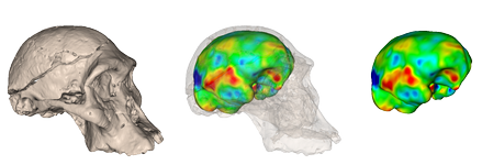
A cranial endocast is a cast (replica) of the inside of the skull. Natural (fossil) endocasts can sometimes be found, and artificial endocasts can be typically obtained by filling the empty braincase with latex and using the latter as a mould to build a plaster cast. More recently, CT has been extensively used to build “virtual” or “digital” endocasts in a precise and non-invasive way.
Studying endocasts is of the utmost importance, because they constitute the only way to indirectly assess global (size, shape) and sometimes local characteristics (meningeal/sulcal/gyral patterns) of the external cerebral anatomy of extinct species, and especially hominids. Due to the difficulty in observing and analysing them before the advent of CT, cranial endocasts have been somewhat neglected in many phylogenetic studies compared to the external skull anatomy, whereas they provide a unique way to look at the evolution of the human brain, and to compare it with that of its closest relatives (i.e. the other primates). In particular, they are key to the understanding of the timing and mode of major morphological changes that have affected the brain during the evolution (in particular the link between brain reorganisation and size increase), and that have supposedly given modern humans the ability for high level cognitive functions.
To date, there exist only a few tools to study virtual endocasts in an automated or semi-automated way. Compared to the study of ectocranial data, that of endocasts poses specific problems, most notably due to the difficulty in defining relevant anatomical landmarks on them. The lack of such objective, reproducible methods, together with the difficulty in accessing the data and interpreting them, partly explain why “paleoneurologists” are still relatively few among physical anthropologists nowadays.
Objectives
The aim of this two-year Collaborative Research Initiative (ARC) granted by INRIA (the French National Institute for Research in Computer Science and Control) is to devise, implement, validate and apply new mathematical and computational methods for the automated morphometric analysis of extant and extinct hominid cranial virtual endocasts. Pre-processing tasks for such an analysis sometimes include automated digital reconstruction of the skull from fragments, correction of taphonomic (i.e. post mortem) deformations, and segmentation of the endocast from CT data. Finally the morphometric analysis of endocasts will be performed over different populations of subjects, including: modern humans, fossil pre-humans and humans (such as South African australopithecines, European Cro-Magnons and Neanderthals), and the closest extant relatives of modern humans (i.e. chimps and bonobos). Of particular interest will be the quantification of asymmetries, as a potential key indicator of brain specialisation.
Participants
This work will be led within a French-Belgian consortium comprising the Visages team at INRIA, Rennes, the Asclepios team at INRIA, Sophia Antipolis, the ICAR team at CNRS, Montpellier, the laboratory of paleoanthropology and anatomical imaging at the Université Paul Sabatier, Toulouse, the Muséum national d’Histoire naturelle at CNRS, Paris, and the laboratory of general histology, neuroanatomy and neuropathology at the Université Libre de Bruxelles.
Symposium
We organised a podium symposium at the 79th annual meeting of the AAPA (American Association of Physical Anthropologists) in Albuquerque, April 2010. The aim of this symposium was to foster the use of computational methods for the automated and objective analysis of virtual hominid endocasts. The talks provided both methodological and applicative views on this problem, with the input from both computer scientists and physical anthropologists.

